REVIEW ARTICLE


Vermamoeba vermiformis - A Free-Living Amoeba with Public Health and Environmental Health Significance


Patrick L. Scheid*
Laboratory of Medical Parasitology, Central Military Hospital Koblenz, Andernacherstr. 100, 56070 Koblenz, Germany


Article Metrics

CrossRef Citations:
0
Total Statistics:

Full-Text HTML Views: 3948
Abstract HTML Views: 788
PDF Downloads: 804
ePub Downloads: 555
Total Views/Downloads: 6095
Unique Statistics:

Full-Text HTML Views: 2229
Abstract HTML Views: 528
PDF Downloads: 531
ePub Downloads: 354
Total Views/Downloads: 3642


Creative Commons License
© 2019 Patrick L. Scheid.

open-access license: This is an open access article distributed under the terms of the Creative Commons Attribution 4.0 International Public License (CC-BY 4.0), a copy of which is available at: (https://creativecommons.org/licenses/by/4.0/legalcode). This license permits unrestricted use, distribution, and reproduction in any medium, provided the original author and source are credited.

* Address correspondence to this author at the Laboratory of Medical Parasitology, Central Military Hospital Koblenz, Andernacherstr. 100, 56070 Koblenz, Germany;
Email: pscheidmedbw@aol.com


Abstract

Many case reports emphasize the fact that Free-Living Amoebae (FLA) can relatively easily get in contact with humans or animals. The presence of several facultative parasitic FLA in habitats related to human activities supports their public health relevance. While some strains of Acanthamoeba, Naegleria fowleri, Balamuthia mandrillaris and several other FLA have been described as facultative human pathogens, it remains controversial whether Vermamoeba vermiformis strains may have a pathogenic potential, or whether this FLA is just an incidental contaminant in a range of human cases. However, several cases support its role as a human parasite, either as the only etiological agent, or in combination with other pathogens. Additionally, a wide range of FLA is known as vectors of microorganisms (endocytobionts), hereby emphasizing their environmental significance. Among those FLA serving as hosts for and vectors of (pathogenic) endocytobionts, there are also descriptions of V. vermiformis as a vehicle and a reservoir of those endocytobionts. The involvement in animal and human health, the role as vector of pathogenic microorganisms and the pathogenicity in cell cultures, led to the assumption that V. vermiformis should be considered relevant in terms of public health and environmental health.

Keywords: Vermamoeba vermiformis, Free-living amoebae, FLA, Endocytobionts, Endosymbionts, Water related parasites, Public Health, Environmental Health.


1. INTRODUCTION

Free-living amoebae (FLA) are predatory heterotrophic protozoa, feeding as trophozoites on bacteria, cyanobacteria, fungi and algae through phagocytosis while adhering to surfaces [1]. There are numerous case reports indicating how easily humans may get in contact with FLA. Some Acanthamoeba sp. strains, Naegleria fowleri, Balamuthia mandrillaris and several other FLA have proved to be facultative human pathogenic microorganisms [2].

The presence of pathogenic FLA in habitats related to human activities supports the public health relevance of these protozoa.

While those FLA genera or strains are well-known parasites of humans and animals, the discussion about the public health and environmental health relevance of V. vermiformis is still ongoing.

Vermamoeba vermiformis was described in 1967 by Page as Hartmannella vermiformis [3, 4]. Smirnov et al. and Page renamed this FLA as Vermamoeba vermiformis for both, morphological and phylogenetic reasons [4]. The genus Echinamoeba is a sister group of Vermamoeba [4-10]. V. vermiformis is widespread in nature and in artificial environments. It is an ubiquitous, cyst-forming FLA, showing a certain resistance (tenacity) in different environmental conditions [11, 12].

In this review study, the impact of V. vermiformis on humans and animals was analysed. An additional systematic literature search was done using databases such as PubMed, but also by screening conference papers of meetings dealing with FLA (e.g. such as the “International Meetings on the biology and pathology of free-living amoebae”). The key words used within the literature search were: “Vermamoeba vermiformis”, “Hartmannella vermiformis”. Those relevant publications dealing with V. (H.) vermiformis were included in this review.

2. MORPHOLOGY AND TAXONOMY

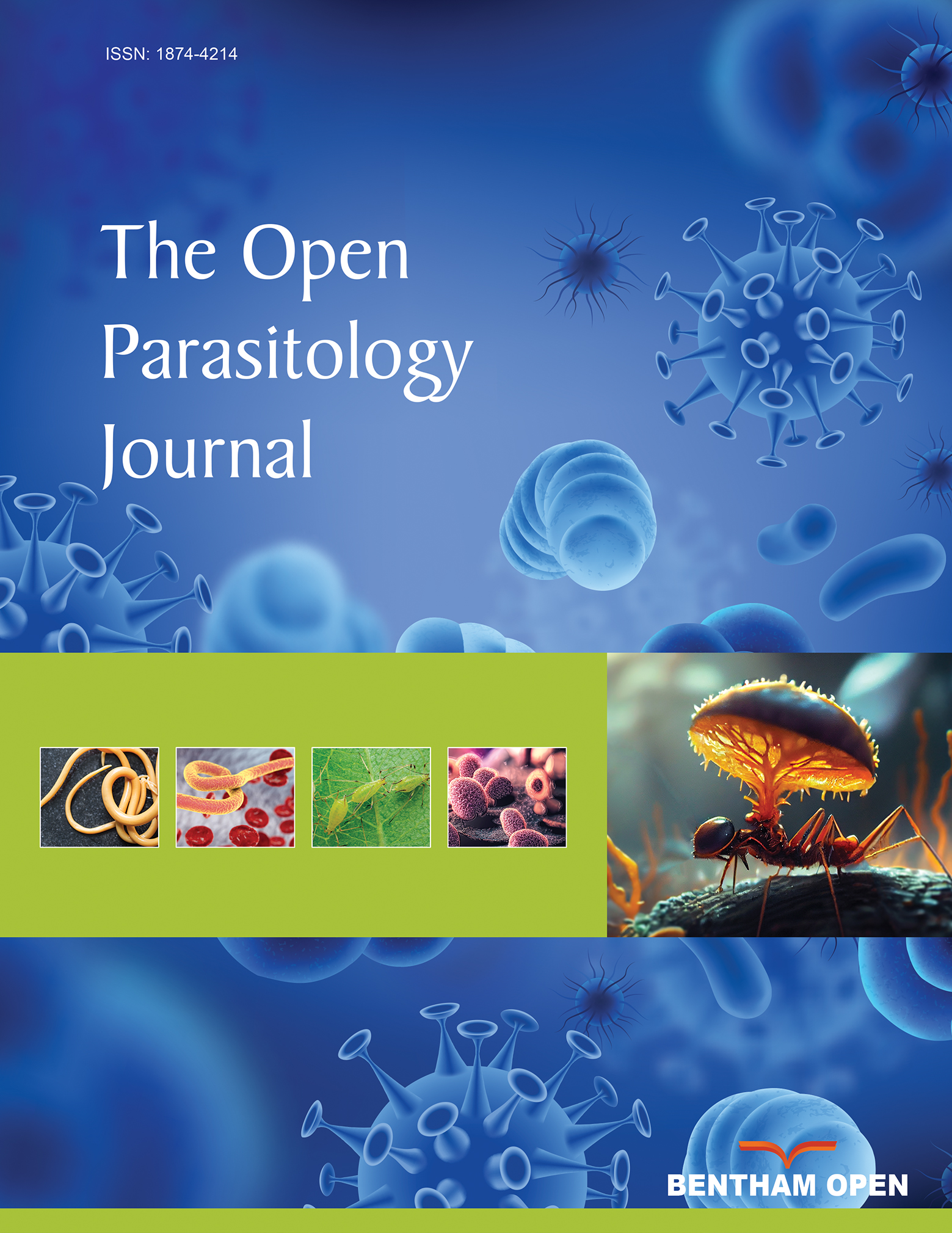
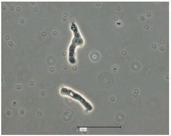
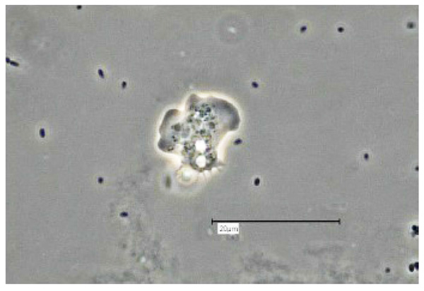
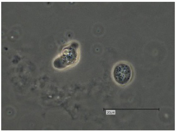
Vermamoeba vermiformis shows a two-stage life cycle with a trophozoite (division by binary fission, feeding, motility) and a dormant cyst form that enables V. vermiformis to survive in hostile environmental conditions such as nutrient depletion, osmotic stress, temperature changes, or pH variation [10]. V. vermiformis can excyst again as soon as the environmental conditions become favourable again. Page and Smirnov et al. (2011) provided morphological descriptions of the trophozoites and cysts of V. vermiformis [4, 13, 14]. These descriptions include morphological features (e.g. length, cylindrical trophozoites, branching with numerous pseudopodia under certain conditions, cyst diameter, and bilayer cyst walls; limax or slug-like motility). A flagellate form has not been described. The trophozoites of V. vermiformis show the typical worm-shaped or slug-like (elongated, cylindrical) morphology described by Page and Smirnov et al. [4, 13]. The locomotive forms are 22-42 μm long. Usually the trophozoites show a monopodial form (Fig. 1) in locomotion, some of them with uroidal filaments (Fig. 2). These uroidal filaments weren`t reported in early studies [3]. Granulae are visible within the cytoplasm as well as a small anterior hyaline zone [10, 15]. In general, morphological features seem to vary among the strains, determined by the environmental conditions. The trophozoites are usually monopodial. However, they may show several pseudopodia and irregular shapes, especially when moving in another direction (Fig. 3).

Fig. (1). V. vermiformis trophozoites, showing the typical monopodial morphology; amoebal saline; Bar: 20µm; phase contrast.

Fig. (2). V. vermiformis trophozoite with prominent uroid filaments; amoebal saline; Bar: 20µm; phase contrast.

Fig. (3). V. vermiformis trophozoite in amoebal saline; showing numerous pseudopodia; Bar: 20µm; phase contrast.

The V. vermiformis cysts are of spherical, round shape with a diameter of 6-9μm. The cyst consists of a 50 nm thick endocyst and a 110 - 140nm thick ectocyst. The two-layered cyst wall (double-wall) contains proteins and glucose polymers. The cyst shows one or two nuclei; no pores or ostioles are visible (Fig. 4) [16]. This cyst stage protects V. vermiformis against hostile environmental conditions such as nutrient depletion, osmotic stress, temperature changes, or pH variation. Fouque et al. (2012) showed that the encystment process of V. vermiformis lasts about 9 hours [11].

V. vermiformis belongs taxonomically to the class Echinamoebida, phylum Tubulinea [4-9].

The phylogenetic discrepancy and the detected morphological differences from the other Hartmannella spp. has led to the introduction of the new genus Vermamoeba, with the only species V. vermiformis. As there is a certain variability in trophozoite morphology and a morphological similarity to other FLA cysts, alternative methods have to be used for accurate detection, including molecular techniques.

Molecular-based analyses of V. vermiformis are limited to sequencing of the 18S rRNA gene, which is used as a phylogenetic marker for taxonomic inferences [10]. Those 18S rRNA sequences, publicly available, show a high degree of sequence conservation for V. vermiformis. This leads to the assumption that the 18S rRNA gene sequences may be insufficient to compare those strains of V. vermifomis with different virulence patterns.

Fig. (4). V. vermiformis trophozoite and cyst; Bar: 20µm; phase contrast.

3. EPIDEMIOLOGY

3.1. Habitats of Vermamoeba vermiformis

Natural freshwater environments, surface water, soil and biofilms are the natural habitats for V. vermiformis. It is one of the most prevalent FLA found in humid ecosystems, other than Acanthamoeba spp [17, 18]. Within its biocenosis, V. vermiformis contributes to microbial communities in biofilms while feeding on bacteria. It has been detected in waters, springs, snow, and soil [19-21].

In addition to these natural humid habitats, V. vermiformis has been isolated from man-made environments and engineered water systems such as tap water, fountains, water pipes, bottled mineral water, drinking water sources, recreational waters and swimming pools [10, 11, 19, 22-28].

In several studies, V. vermiformis has been found within the human environment, showing paradigmatically how easily humans may find themselves in close contact with these FLA. V. vermiformis has been detected in composts, aerosols from composting facilities, wastewater from the textile industry [10, 28, 29], contact lens storage cases, and even hospital ward dust and biofilms [24, 30-33].

V. vermiformis has been found in thermal springs with temperatures of 41-53 °C and a pH of 4.9-7 [27, 34-36]. Rohr et al. identified V. (resp. Hartmannella) vermiformis in 65% of the hot water samples which were positive for amoebae and on 15% of swabs from moist areas [37, 38].

3.2. Vermamoeba vermiformis as a Parasite for Humans

The role of V. vermiformis as a potential pathogenic parasite of humans has been discussed for many years. Some authors have concluded that V. vermiformis is an environmental contaminant without any involvement in pathogenesis [39-41].

A certain cytopathogenicity of the thermo-tolerant V. vermiformis on keratocytes was confirmed in in vitro studies [41, 42]. Another hint regarding the (veterinary medical) pathogenic potential is the fact that some strains of V. (Hartmannella) vermiformis have produced tissue lesions in experimentally infected fish [15]. At least, pathogenicity of V. vermiformis for aquatic organisms cannot be excluded.

The medical significance of V. vermiformis (resp. Hartmannella vermiformis) and especially its pathogenic potential as a parasite for humans has been reported from several countries, including Spain, Costa Rica, Peru, France, Scotland, Japan and Iran [16, 43, 46-49]. The isolation of V. vermiformis from humans or while examining human infections, has provided evidence of its potential involvement in the pathogenesis. In 2014, V. vermiformis was detected in human nasal swabs [16]. Most of the “case reports” with an involvement of V. vermiformis have included corneal damage [10]. A keratitis case included a female contact lens wearer, who presented initially with eye pain, redness, blurred vision, photosensitivity, tearing, and a sensation of a foreign body in her eye [45]. In other cases V. vermiformis was isolated while examining for an Acanthamoeba keratitis [50-53]. Interestingly, V. vermiformis has been found predominantly in mixed amoebic keratitis cases [46, 50, 51, 53, 54]. These findings, together with the results of other studies, have led to the conclusion that the spectrum of protozoa contaminating contact lens solutions is broader than previously known and includes Acanthamoeba spp., Balamuthia mandrillaris and also V. vermiformis [44, 50-53].

Two human cases involving V. vermiformis have been recently examined in Germany: V. vermiformis was detected within the contact lens cases of a bacterial keratitis patient. In this “case report” V. vermiformis seemed to be a contaminant without (significant) involvement in the pathogenesis.

A second case included the only non-keratitis case report so far - with an exclusive isolation of V. vermiformis from a human. V. vermiformis has been confirmed as potential etiological agent in a 27 years old female patient, who presented with a weeping wound developing as a painful ulcer on the upper eyelid at the medial angle of the right eye. Cultivation, morphological microscopical analysis and nucleic acid techniques, followed by sequence analysis revealed V. vermiformis as the only detected microorganism involved in this case. The confirmed presence of V. vermiformis in the ulcer and the proven absence of the typical pathogenic bacterial microorganisms led to the strong assumption that V. vermiformis may be involved in the pathogenesis of this human case.

3.3. Vermamoeba vermiformis as Vector of Endocytobionts

In general, naked free-living amoebae (FLA) graze in biofilms and feed on bacteria, algae, yeasts and other protozoa. They capture their prey by phagocytosis following chemotactic trophism and transfer them to lysosomal compartments in the phagocytic pathway where they are usually digested by enzymes [1]. However, some of these intracellular microorganisms (endocytobionts) have developed a strategy to avoid lysis and digestion during the phagocytic process. The impact of such interactions of FLA and their endocytobionts with respect to Public Health and Environmental Health was described recently [54, 55]. Additionally, a wide range of FLA is known to be vectors of endocytobionts [2].

The relevance of the FLA - endocytobiont relationship in terms of pathogenicity, tenacity, virulence enhancement, protection, gene transfer etc. is the focus of current research [56].

While there is still an ongoing discussion about the public health relevance of V. vermiformis as a human parasite, it has proven to be a vector of endocytobionts, some of these being confirmed pathogens of public health significance [for overview see: 10 or 57]. These endocytobionts may also play a significant role in aggravating the infection or in enhancing inflammatory processes [14, 58].

In 1988 V. (Hartmannella) vermiformis was described as an important protozoon in the ecology of Legionella pneumophila [59]. Since then V. vermiformis is known as a reservoir and vector of Legionellae [17, 60-64].

V. vermiformis also has proven to serve as a vehicle for and vector of Bacillus anthracis, Neochlamydia hartmannellae and other Chlamydia-like endocytobionts [65-67; for overview see: 10, 57 or 60]. Other bacterial microorganisms persisting as endocytobionts and replicating intracellularly in V. vermiformis are Protochlamydia massiliensis, Protochlamydia phocaeensis and Rubidus massiliensis [68-70].

V. vermiformis may also be associated with Mycobacteriae. in vitro studies revealed that highly pathogenic Mycobacterium leprae remained viable and virulent within V. vermiformis cysts [10, 71, 79]. Even on nasal swabs V. vermiformis and Mycobacterium chelonae were detected sympatrically [16].

V. vermiformis can be permissive for Pseudomonas aeruginosa [72]. There are also interesting relationships of V. vermiformis with Stenotrophomonas maltophilia (an important nosocomial pathogen) and Campylobacter jejuni [19, 33, 73-75].

Francisella novicida has proven to be another endocytobiont of V. vermiformis, surviving and replicating intracellularly in non-acidified phagosomes [76].

An intranuclear endocytobiont of V. vermiformis has been described recently and named Nucleicultrix amoebiphila [77].

As V. vermiformis is isolated frequently in environmental samples, it has been used as a potential host in the search for giant viruses (giruses). Some of those giruses were detected in V. vermiformis: Several isolates of Faustovirus, an Asfarvirus-related lineage of giruses, were isolated using V. vermiformis as the host amoeba [78]. Kaumoebavirus and Orpheovirus are further examples of giruses proliferating in V. vermiformis [79, 80].

Interactions between V. vermiformis and fungi include Exophiala dermatitidis, Aspergillus fumigatus, Candida spp. and Fusarium oxysporum [14, 81-84].

In the context of acting as vector of pathogenic endocytobionts, V. vermiformis may also play an important role in the distribution of food borne microorganisms. Several species of FLA have been discovered on vegetables, green salad, spinach, lettuce, as well as in chicken coops and refrigerators. Studies to determine the abundance of free-living protozoa on dishcloths as a possible source for cross-contamination in food processing establishments have been conducted recently. Vahlkampfia, Vannella, Acanthamoeba, Hyperamoeba, and Vermamoeba have been detected on these dishcloths [53, 55].

CONCLUSION

V. vermiformis is certainly another FLA to be considered as a potential parasite of humans or animals and as a vector of encocytobionts. The case reports with an involvement of V. vermiformis and especially one of the recent cases with V. vermiformis as the only detected microorganism, indicate that this FLA may be potentially (or opportunistically) pathogenic. The thermo-tolerant V. vermiformis should definitely be considered in future studies and/or diagnostics targeting FLA as etiological agents of human and animal diseases. When comparing the studies of V. vermiformis as a potential parasite for humans and its role as vector of potentially pathogenic microorganisms, we must (still) come to the conclusion that V. vermifomis is more important as a vector, according to the literature available at present.

CONSENT FOR PUBLICATION

Not applicable.

FUNDING

Declared none.

CONFLICT OF INTEREST

The authors declare no conflict of interest, financial or otherwise.

ACKNOWLEDGEMENTS

The author would like to thank Dr. David Lam (MD, MPH, Shaman Medical Consulting) for review and English language editing of the article and Dr. C. Balczun for fruitful discussions.

REFERENCES

[1] Scheid P. Free-living amoebae and their multiple impacts on environmental health.
[2] Scheid P. Lebensmittelassoziierte parasiten: Helminthen und protozoen 2018.
[3] Page FC. Taxonomic criteria for limax amoebae, with descriptions of 3 new species of Hartmannella and 3 of Vahlkampfia. J Protozool 1967; 14(3): 499-521.
[4] Smirnov AV, Chao E, Nassonova ES, Cavalier-Smith T. A revised classification of naked lobose amoebae (Amoebozoa: lobosa). Protist 2011; 162(4): 545-70.
[5] Bolivar I, Fahrni JF, Smirnov A, Pawlowski J. SSU rRNA-based phylogenetic position of the genera Amoeba and Chaos (Lobosea, Gymnamoebia): The origin of gymnamoebae revisited. Mol Biol Evol 2001; 18(12): 2306-14.
[6] Corsaro D, Michel R, Walochnik J, Müller KD, Greub G. Saccamoeba lacustris, sp. nov. (Amoebozoa: Lobosea: Hartmannellidae), a new lobose amoeba, parasitized by the novel chlamydia ‘Candidatus Metachlamydia lacustris’ (Chlamydiae: Parachlamydiaceae). Eur J Protistol 2010; 46(2): 86-95.
[7] Brown MW, Silberman JD, Spiegel FW. “Slime molds” among the Tubulinea (Amoebozoa): Molecular systematics and taxonomy of Copromyxa. Protist 2011; 162(2): 277-87.
[8] Watson PM, Sorrell SC, Brown MW. Ptolemeba n. gen., a novel genus of hartmannellid amoebae (Tubulinea, Amoebozoa); with an emphasis on the taxonomy of Saccamoeba. J Eukaryot Microbiol 2014; 61(6): 611-9.
[9] Adl SM, Bass D, Lane CE, et al. Revision to the classification, nomenclature, and diversity of eukaryotes. J Eukaryot Microbiol 2019; 66(1): 4-119.
[10] Delafont V, Rodier M-H, Maisonneuve E, Cateau E. Vermamoeba vermiformis: A free-living amoeba of interest. Microb Ecol 2018; 76(4): 991-1001.
[11] Fouque E, Héchard Y, Hartemann P, Humeau P, Trouilhé MC. Sensitivity of Vermamoeba (Hartmannella) vermiformis cysts to conventional disinfectants and protease. J Water Health 2015; 13(2): 302-10.
[12] Fouque E, Yefimova M, Trouilhé M-C, et al. Morphological study of the encystment and excystment of Vermamoeba vermiformis revealed original traits. J Eukaryot Microbiol 2015; 62(3): 327-37.
[13] Page F. Nackte Rhizopoda und Heliozoa, Protozoenfauna 1991; Vol. 2
[14] Masangkay F, Milanez G, Karanis P, Nissapatorn V. Vermamoeba vermiformis—global trend and future perspective2018.
[15] Dyková I, Pindová Z, Fiala I, Dvoráková H, Machácková B. Fish-isolated strains of Hartmannella vermiformis page, 1967: Morphology, phylogeny and molecular diagnosis of the species in tissue lesions. Folia Parasitol (Praha) 2005; 52(4): 295-303.
[16] Cabello-Vílchez A, Mena R, Zuñiga J, et al. Reyes-Batlle M, Piñero J, Valladares B, Lorenzo-Morales J. Endosymbiotic Mycobacterium chelonae in a Vermamoeba vermiformis strain isolated from the nasal mucosa of an HIV patient in Lima, Peru. Exp Parasitol 2014; 145(Suppl.): 127-30.
[17] Hsu BM, Lin CL, Shih FC. Survey of pathogenic free-living amoebae and Legionella spp. in mud spring recreation area. Water Res 2009; 43(11): 2817-28.
[18] Wang H, Edwards M, Falkinham JO III, Pruden A. Molecular survey of the occurrence of Legionella spp., Mycobacterium spp., Pseudomonas aeruginosa, and amoeba hosts in two chloraminated drinking water distribution systems. Appl Environ Microbiol 2012; 78(17): 6285-94.
[19] Cateau E, Delafont V, Hechard Y, Rodier MH. Free-living amoebae: What part do they play in healthcare-associated infections? J Hosp Infect 2014; 87(3): 131-40.
[20] Valster RM, Wullings BA, Bakker G, Smidt H, van der Kooij D. Free-living protozoa in two unchlorinated drinking water supplies, identified by phylogenic analysis of 18S rRNA gene sequences. Appl Environ Microbiol 2009; 75(14): 4736-46.
[21] Thomas V, Loret JF, Jousset M, Greub G. Biodiversity of amoebae and amoebae-resisting bacteria in a drinking water treatment plant. Environ Microbiol 2008; 10(10): 2728-45.
[22] Bullerwell CE, Burger G, Gott JM, Kourennaia O, Schnare MN, Gray MW. Abundant 5S rRNA-like transcripts encoded by the mitochondrial genome in amoebozoa. Eukaryot Cell 2010; 9(5): 762-73.
[23] Anderson OR. The role of amoeboid protists and the microbial community in moss-rich terrestrial ecosystems: Biogeochemical implications for the carbon budget and carbon cycle, especially at higher latitudes. J Eukaryot Microbiol 2008; 55(3): 145-50.
[24] Kuiper MW, Valster RM, Wullings BA, Boonstra H, Smidt H, van der Kooij D. Quantitative detection of the free-living amoeba Hartmannella vermiformis in surface water by using real-time PCR. Appl Environ Microbiol 2006; 72(9): 5750-6.
[25] Armand B, Motazedian MH, Asgari Q. Isolation and identification of pathogenic free-living amoeba from surface and tap water of Shiraz City using morphological and molecular methods. Parasitol Res 2016; 115(1): 63-8.
[26] Nazar M, Haghighi A, Taghipour N, et al. Molecular identification of Hartmannella vermiformis and Vannella persistens from man-made recreational water environments, Tehran, Iran. Parasitol Res 2012; 111(2): 835-9.
[27] Montalbano Di Filippo M, Santoro M, Lovreglio P, et al. Isolation and molecular characterization of free-living amoebae from different water sources in Italy. Int J Environ Res Public Health 2015; 12(4): 3417-27.
[28] Ramirez E, Robles E, Martinez B, et al. Distribution of free-living amoebae in a treatment system of textile industrial wastewater. Exp Parasitol 2014; 145(Suppl.): S34-8.
[29] Conza L, Pagani SC, Gaia V. Presence of Legionella and free-living Amoebae in composts and bioaerosols from composting facilities. PLoS One 2013; 8(7): e68244.
[30] Thomas V, Herrera-Rimann K, Blanc DS, Greub G. Biodiversity of amoebae and amoeba-resisting bacteria in a hospital water network. Appl Environ Microbiol 2006; 72(4): 2428-38.
[31] Lasjerdi Z, Niyyati M, Haghighi A, et al. Potentially pathogenic free-living amoebae isolated from hospital wards with immunodeficient patients in Tehran, Iran. Parasitol Res 2011; 109(3): 575-80.
[32] Lasjerdi Z, Niyyati M, Lorenzo-Morales J, Haghighi A, Taghipour N. Ophthalmology hospital wards contamination to pathogenic free living Amoebae in Iran. Acta Parasitol 2015; 60(3): 417-22.
[33] Pagnier I, Valles C, Raoult D, La Scola B. Isolation of Vermamoeba vermiformis and associated bacteria in hospital water. Microb Pathog 2015; 80: 14-20.
[34] Gianinazzi C, Schild M, Zumkehr B, et al. Screening of Swiss hot spring resorts for potentially pathogenic free-living amoebae. Exp Parasitol 2010; 126(1): 45-53.
[35] Solgi R, Niyyati M, Haghighi A, Mojarad EN. Occurrence of thermotolerant Hartmannella vermiformis and Naegleria spp. in hot springs of Ardebil Province, Northwest Iran. Iran J Parasitol 2012; 7(2): 47-52.
[36] Rhoads WJ, Ji P, Pruden A, Edwards MA. Water heater temperature set point and water use patterns influence Legionella pneumophila and associated microorganisms at the tap. Microbiome 2015; 3: 67.
[37] Rohr U, Weber S, Michel R, Selenka F, Wilhelm M. Comparison of free-living amoebae in hot water systems of hospitals with isolates from moist sanitary areas by identifying genera and determining temperature tolerance. Appl Environ Microbiol 1998; 64(5): 1822-4.
[38] Cateau E, Imbert C, Rodier MH. Hartmanella vermiformis can be permissive for Pseudomonas aeruginosa. Lett Appl Microbiol 2008; 47(5): 475-7.
[39] De Jonckheere JF, Brown S. There is no evidence that the free-living ameba Hartmannella is a human parasite. Clin Infect Dis 1998; 26(3): 773.
[40] De Jonckheere JF, Brown S. Is the free-living ameba Hartmannella causing keratitis? Clin Infect Dis 1998; 27(5): 1337-8.
[41] Kinnear FB. Non-Acanthamoeba amoebic keratitis. J Infect 2001; 42(3): 218-9.
[42] Kinnear FB. Cytopathogenicity of acanthamoeba, vahlkampfia and hartmannella: Quantative & qualitative in vitro studies on keratocytes. J Infect 2003; 46(4): 228-37.
[43] Lorenzo-Morales J, Martínez-Carretero E, Batista N, et al. Early diagnosis of amoebic keratitis due to a mixed infection with Acanthamoeba and Hartmannella. Parasitol Res 2007; 102(1): 167-9.
[44] Bouchoucha I, Aziz A, Hoffart L, Drancourt M. Repertoire of free-living protozoa in contact lens solutions. BMC Ophthalmol 2016; 16(1): 191.
[45] Abedkhojasteh H, Niyyati M, Rahimi F, Heidari M, Farnia S, Rezaeian M. First report of Hartmannella keratitis in a cosmetic soft contact lens wearer in Iran. Iran J Parasitol 2013; 8(3): 481-5.
[46] Inoue T, Asari S, Tahara K, Hayashi K, Kiritoshi A, Shimomura Y. Acanthamoeba keratitis with symbiosis of Hartmannella ameba. Am J Ophthalmol 1998; 125(5): 721-3.
[47] Walochnik J, Scheikl U, Haller-Schober EM. Twenty years of acanthamoeba diagnostics in Austria. J Eukaryot Microbiol 2015; 62(1): 3-11.
[48] Kennedy SM, Devine P, Hurley C, Ooi YS, Collum LM. Corneal infection associated with Hartmannella vermiformis in contact-lens wearer. Lancet 1995; 346(8975): 637-8.
[49] Aitken D, Hay J, Kinnear FB, Kirkness CM, Lee WR, Seal DV. Amebic keratitis in a wearer of disposable contact lenses due to a mixed Vahlkampfia and Hartmannella infection. Ophthalmology 1996; 103(3): 485-94.
[50] De Jonckheere JF, Brown S. Non-Acanthamoeba amoebic keratitis. Cornea 1999; 18(4): 499-501.
[51] Scheid P, Zöller L, Pressmar S, Richard G, Michel R. An extraordinary endocytobiont in Acanthamoeba sp. isolated from a patient with keratitis. Parasitol Res 2008; 102(5): 945-50.
[52] Gray TB, Cursons RT, Sherwan JF, Rose PR. Acanthamoeba, bacterial, and fungal contamination of contact lens storage cases. Br J Ophthalmol 1995; 79(6): 601-5.
[53] Balczun C, Scheid PL. Detection of Balamuthia mandrillaris DNA in the storage case of contact lenses in Germany. Parasitol Res 2016; 115(5): 2111-4.
[54] Balczun C, Scheid PL. Free-living amoebae as hosts for and vectors of intracellular microorganisms with public health significance. Viruses 2017; 9(4): 65.
[55] Scheid P. Free-living amoebae and their multiple impacts on environmental health. Reference Module in Earth Systems and Environmental Sciences 2018.
[56] Scheid P. Viruses in close associations with free-living amoebae. Parasitol Res 2015; 114(11): 3959-67.
[57] Scheid P. Relevance of free-living amoebae as hosts for phylogenetically diverse microorganisms. Parasitol Res 2014; 113(7): 2407-14.
[58] Muchesa P, Leifels M, Jurzik L, Hoorzook KB, Barnard TG, Bartie C. Coexistence of free-living amoebae and bacteria in selected South African hospital water distribution systems. Parasitol Res 2017; 116(1): 155-65.
[59] Wadowsky RM, Butler LJ, Cook MK, et al. Growth-supporting activity for Legionella pneumophila in tap water cultures and implication of hartmannellid amoebae as growth factors. Appl Environ Microbiol 1988; 54(11): 2677-82.
[60] Fields BS, Nerad TA, Sawyer TK, et al. Characterization of an axenic strain of Hartmannella vermiformis obtained from an investigation of nosocomial legionellosis. J Protozool 1990; 37(6): 581-3.
[61] Brieland JK, Fantone JC, Remick DG, LeGendre M, McClain M, Engleberg NC. The role of Legionella pneumophila-infected Hartmannella vermiformis as an infectious particle in a murine model of Legionnaire’s disease. Infect Immun 1997; 65(12): 5330-3.
[62] Garcia A, Goñi P, Cieloszyk J, et al. Identification of free-living amoebae and amoeba-associated bacteria from reservoirs and water treatment plants by molecular techniques. Environ Sci Technol 2013; 47(7): 3132-40.
[63] Tyson JY, Pearce MM, Vargas P, Bagchi S, Mulhern BJ, Cianciotto NP. Multiple Legionella pneumophila Type II secretion substrates, including a novel protein, contribute to differential infection of the amoebae Acanthamoeba castellanii, Hartmannella vermiformis, and Naegleria lovaniensis. Infect Immun 2013; 81(5): 1399-410.
[64] Kuiper MW, Wullings BA, Akkermans AD, Beumer RR, van der Kooij D. Intracellular proliferation of Legionella pneumophila in Hartmannella vermiformis in aquatic biofilms grown on plasticized polyvinyl chloride. Appl Environ Microbiol 2004; 70(11): 6826-33.
[65] Dey R, Hoffman PS, Glomski IJ. Germination and amplification of anthrax spores by soil-dwelling amoebas. Appl Environ Microbiol 2012; 78(22): 8075-81.
[66] Horn M, Wagner M, Müller K-D, et al. Neochlamydia hartmannellae gen. nov., sp. nov. (Parachlamydiaceae), an endoparasite of the amoeba Hartmannella vermiformis. Microbiology 2000; 146(Pt 5): 1231-9.
[67] Henning K, Zöller L, Hauroeder B, Hotzel H, Michel R. Hartmannella vermiformis (Hartmannellidae) harboured a hidden Chlamydia-like endosymbiont. Endocytobiosis Cell Res 2007; 18: 1-10.
[68] Benamar S, Bou Khalil JY, Blanc-Tailleur C, Bilen M, Barrassi L, La Scola B. Developmental cycle and genome analysis of Protochlamydia massiliensis sp. nov. A new species in the Parachlamydiacae family. Front Cell Infect Microbiol 2017; 7: 385.
[69] Bou KJY, Benamar S, Di Pinto F, Blanc-Tailleur C, Raoult D, La Scola B. Protochlamydia phocaeensis sp. nov., a new Chlamydiales species with host dependent replication cycle. Microbes Infect 2017; 19(6): 343-50.
[70] Bou KJY, Benamar S, Baudoin JP, et al. Developmental cycle and genome analysis of Rubidus massiliensis, a new Vermamoeba vermiformis pathogen. Front Cell Infect Microbiol 2016; 6: 31.
[71] Wheat WH, Casali AL, Thomas V, et al. Long-term survival and virulence of Mycobacterium leprae in amoebal cysts. PLoS Negl Trop Dis 2014; 8(12): e3405.
[72] Cateau E, Imbert C, Rodier MH. Hartmanella vermiformis can be permissive for Pseudomonas aeruginosa. Lett Appl Microbiol 2008; 47(5): 475-7.
[73] Evstigneeva A, Raoult D, Karpachevskiy L, La Scola B. Amoeba co-culture of soil specimens recovered 33 different bacteria, including four new species and Streptococcus pneumoniae Microbiology 2009; 155: 657-64.
[74] Axelsson-Olsson D, Olofsson J, Svensson L, et al. Amoebae and algae can prolong the survival of Campylobacter species in co-culture. Exp Parasitol 2010; 126(1): 59-64.
[75] Denet E, Vasselon V, Burdin B, Nazaret S, Favre-Bonté S. Survival and growth of Stenotrophomonas maltophilia in free-living amoebae (FLA) and bacterial virulence properties. PLoS One 2018; 13(2): e0192308.
[76] Santic M, Ozanic M, Semic V, Pavokovic G, Mrvcic V, Kwaik YA. Intra-vacuolar proliferation of F. novicida within H. vermiformis. Front Microbiol 2011; 2: 78.
[77] Schulz F, Lagkouvardos I, Wascher F, Aistleitner K, Kostanjšek R, Horn M. Life in an unusual intracellular niche: A bacterial symbiont infecting the nucleus of amoebae. ISME J 2014; 8(8): 1634-44.
[78] Reteno DG, Benamar S, Khalil JB, et al. Faustovirus, an asfarvirus-related new lineage of giant viruses infecting amoebae. J Virol 2015; 89(13): 6585-94.
[79] Bajrai LH, Benamar S, Azhar EI, et al. Kaumoebavirus, a new virus that clusters with Faustoviruses and Asfarviridae. Viruses 2016; 8(11): 8.
[80] Andreani J, Khalil J, Baptiste E, et al. Orpheovirus IHUMI-LCC2: A new virus among the giant viruses. Front Microbiol 2017.
[81] Cateau E, Mergey T, Kauffmann-Lacroix C, Rodier MH. Relationships between free living amoebae and Exophiala dermatitidis: A preliminary study. Med Mycol 2009; 47(1): 115-8.
[82] Maisonneuve E, Cateau E, Kaaki S, Rodier MH. Vermamoeba vermiformis-Aspergillus fumigatus relationships and comparison with other phagocytic cells. Parasitol Res 2016; 115(11): 4097-105.
[83] Vanessa B, Virginie M, Nathalie Q, Marie-Hélène R, Christine I. Hartmannella vermiformis can promote proliferation of Candida spp. in tap-water. Water Res 2012; 46(17): 5707-14.
[84] Chavatte N, Baré J, Lambrecht E, et al. Co-occurrence of free-living protozoa and foodborne pathogens on dishcloths: Implications for food safety. Int J Food Microbiol 2014; 191: 89-96.